
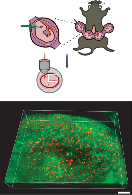
To overcome these limitations and allow imaging in the embryo over multiple hours, researchers in the labs of Zengcai Guo and Da Mi at Tsinghua University in Beijing developed an intravital imaging of externally immobilized embryos (IMEE) approach. The researchers open the uterine wall and carefully lift an embryo out while keeping the amniotic sac intact. The embryo is pulled through a thin film at the bottom of an imaging chamber. One can then rotate the embryo to achieve a desired field of view before gently immobilizing it with agarose and suction within the device. The proper temperature can be maintained by pumping warm water through the device.
The researchers could image embryos within an age range of E10.5 to E16.5. Using a variety of methods such as blood flow assessment or gene expression analysis, the researchers determined that they could maintain the embryos in an externally immobilized state for up to 8 hours without deleterious effects such as inflammation or morphological abnormalities.