
Sample collection
Biopsy samples were collected from n = 12 RA-diagnosed individuals at the Orthopedic Department of AIIMS (All India Institute of Medical Sciences), New Delhi, India, who fulfilled the 2010 diagnostic criteria established by the American College of Rheumatology (ACR) and the European League Against Rheumatism (EULAR)3. The medical histories of all patients were recorded (Supplementary Table 1). The study protocol complied with the Declaration of Helsinki, approved by the ethical committee of Council of Scientific & Industrial Research-Institute of Genomics & Integrative Biology (CSIR-IGIB), New Delhi, India, and AIIMS, New Delhi, India, Registration No. IEC-237/07.05.2021, RP-18/2021, confirming that all experiments were performed in accordance with relevant guidelines and regulations. Additionally, all participants gave their informed consent before taking part in the study.
Cell culture (SW982) and TNF-α induction
SW982 cells, obtained from the National Centre for Cell Science (NCCS) in India, are commonly utilized in RA synovitis studies. These cells were cultured at 37 °C in a humidified environment with 5% CO₂, using Dulbecco’s Modified Eagle Medium (DMEM) supplemented with 10% Fetal Bovine Serum (FBS) and antibiotics. To investigate the effect of miR-4693-5p expression under RA mimic conditions, we created an inflammatory state similar to RA by stimulating the SW982 cells with TNF-α (10 ng/ml)12.
SW982, a human synovial sarcoma-derived cell line, is widely used as an in vitro model for RA studies due to its fibroblast-like morphology and similarity to RA-FLS. Like RA-FLS, SW982 cells express pro-inflammatory mediators (TNF-α, IL-6, IL-1β, MMPs) upon cytokine stimulation13. While primary RA-FLS are influenced by complex in vivo factors like immune interactions and prior drug exposure, SW982 cells offer a more controlled environment for investigating cytokine-mediated molecular responses14. Therefore, SW982 cells were used in this study to specifically examine TNF-α-mediated regulation of miR-4693-5p expression under controlled conditions.
Real-time PCR (real-time polymerase chain reaction)
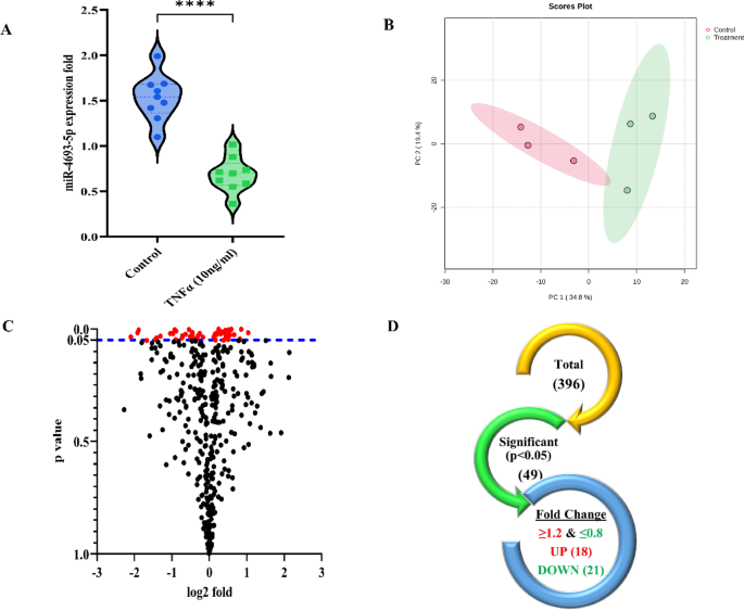
SW982 cells were grown in a six-well plate. After 70–80% confluency, cells were induced with TNFα (10 ng/ml) for 24 h to mimic the RA condition in SW982 cells. Following the manufacturer’s instructions, RNA was extracted using Tri-Xtract (G Biosciences). Afterward, isolated RNA (1 µg) was employed for cDNA synthesis with the cDNA Synthesis Kit (G Biosciences)15. A stem-loop primer was utilized to evaluate the expression of miR-4693-5p in TNF-α induced SW982 cells using a specific primer (Supplementary Table 2) for miR-4693-5p and U6 as an internal control mixed with 5x Hot Fire pol EvaGreen qPCR master mix (Solis BioDyne). miR-4693-5p expression was quantified using the Roche LightCycler 480 II real-time PCR system. The data was evaluated quantitatively using the 2-ΔΔCT formula16.
Primary cell (RAFLS) isolation
To evaluate the physiological function of miR-4693-5p in RA, synovial tissue was carefully minced and digested with 0.5 mg/ml collagenase (Sigma, St. Louis, MO, USA). The digested tissue was cultured in DMEM supplemented with 10% FBS at 37 °C in a 5% CO₂ incubator. Cells were treated after reaching the second passage17.
Cell transfection
To evaluate the function of miR-4693-5p, cells were cultured in a 6-well plate until they reached 60–70% confluency. Transfection was performed with 25 nM of either a miRNA Negative Control (NC) or a miR-4693-5p mimic (Invitrogen, Waltham, MA, USA), along with Lipofectamine RNAiMAX Transfection Reagent (Invitrogen) in Opti-MEM media (Gibco, Waltham, MA, USA). After 5 h, the media was replaced with DMEM supplemented with 10% FBS9.
Preparation of RAFLS for proteomics: SWATH-MS acquisition
RAFLS transfected with miR-4693-5p and NC cells were lysed, and the extracted proteins were pooled and quantified using the Bicinchoninic Acid (BCA) assay. Protein (100 µg) was digested overnight with trypsin (0.1 µg/µl, Promega, USA) at 37 °C, and peptides were extracted using 50% acetonitrile (ACN) and 0.1% trifluoroacetic acid (TFA, Sigma Aldrich, USA). The mixture was dried in a speed-vac, and peptides were separated using an SCX cartridge (5 microns, 300 Å beads, AB Sciex, USA). Elution was performed with 70% v/v acetonitrile and 0.1% formic acid. In SWATH-MS quantitative acquisition, proteins with at least two unique peptides and an unused score of 2.0 or higher were considered, as outlined in previous studies18,19.
Multivariate analysis
Principal component analysis (PCA) was used to identify group differences. PCA can discrete predictive variation from uncorrelated differences to facilitate understanding of different sources of variation. Pareto scaling was used for normalization by a metaboanalyst20.
Construction of PPI (protein-protein interaction) network and enrichment analysis of selected targets
To explore the biological processes, cellular components, and molecular functions of the RA-related proteins, Cytoscape 3.8.2 was utilized21,22. The analysis incorporated Gene Ontology (GO), Kyoto Encyclopedia of Genes and Genomes (KEGG)23, and wiki pathways to study the interactions between connected proteins, aiding in the development of hypotheses and understanding their associated functional pathways24.
Common targets for miR-4693-5p
A detailed analysis was conducted to explore the potential targets of miR-4693-5p and their relevance to the development of RA. The Venny tool was used to compare the predicted targets of miR-4693-5p from miRNA target databases (miRDB, TargetScan, and RNA22) with the SWATH DEPs21. This comparison allowed us to identify potential targets of miR-4693-5p and to understand how they are related to specific proteins involved in the pathogenesis of RA9.
Protein extraction and western blot analysis
RAFLS cells were cultured to 70–80% confluency and then transfected with miR-4693-5p for 48 h. After incubation, cells were lysed on ice using RIPA buffer. Protein concentration was quantified using the BCA method, and 40 µg of protein was loaded onto an SDS-PAGE gel for electrophoretic separation. Proteins were then transferred onto a NC membrane (G Biosciences) via a semi-dry transfer system (Bio-Rad Laboratories). The membrane was blocked with 3% BSA for 2 h and incubated overnight at 4 °C with primary antibodies (Santa Cruz), including Anti-human FN-1 (1:500), E-Cadherin (E-CAD, 1:3000), N-Cadherin (N-CAD, 1:2000), Vascular Endothelial Growth Factor (VEGF, 1:2000), CALD1 (1:2000), YWHAZ (1:1000), SOD2 (1:1000), Catalase (1:1000), Glutathione Peroxidase-1 (GPx1, 1:1000), and Ki67 (1:1000). After three washes with 1X TBST, the membrane was incubated with HRP-conjugated secondary antibodies (1:5000, Jackson ImmunoResearch) for one hour at room temperature. The membrane was washed and treated with enhanced chemiluminescence (ECL) reagent, and protein bands were visualized using the ChemiDoc imaging system and analyzed by Image lab software (Bio-Rad Laboratories)25,26.
MTT assay for cell proliferation assessment
To assess the effect of miR-4693-5p on cell proliferation, an MTT assay was conducted. RA-FLS cells (5,000 per well) were plated in 96-well plates and cultured until reaching 70–80% confluency. The cells were then transfected with 25 nM of miR-4693-5p and incubated for 48 h. For the assay, 10 µl of MTT solution (4 mg/ml) was added to each well, followed by incubation for 3 h to allow the formation of formazan crystals. To dissolve the crystals, 100 µl of dimethyl sulfoxide (DMSO) was added. The absorbance was then measured at 570 nm using a microplate reader to quantify cell proliferation. A 20% DMSO solution was used as a toxic control to validate the assay’s sensitivity27.
Hoechst 33342 staining
Apoptosis and typical morphological features were examined by Hoechst 33342. After transfection, the cells were gently washed with cold 1X PBS twice to remove residual media. Cells were fixed using 4% paraformaldehyde for 30 min at room temperature (RT). After fixation, the cells were rinsed with 1X PBS and stained with 5 µg/ml Hoechst 3334228. The staining was carried out in the dark at 37 °C for 20 min to protect the dye from light degradation. Finally, the cells underwent two additional PBS washes, and apoptotic cells were identified based on nuclear morphology. The relative fluorescence intensity of apoptotic cells was analyzed using a fluorescence Zoe microscope (Bio-Rad Laboratories). Apoptotic cells absorb Hoechst dye more quickly than non-apoptotic cells. Normal cells show organized blue chromatin, while apoptotic cells have bright blue, highly condensed, or fragmented chromatin29,30.
MitoSOX Red‑mitochondrial superoxide production (analysis of mitochondrial superoxide production) in RA
To assess mitochondrial superoxide production and evaluate the effects of miR-4693-5p in RAFLS, we used MitoSOXRed (Thermo Fisher, Monza, IT). The cells were incubated with MitoSOXRed for 30 min and then washed with PBS to remove excess dye. The production of superoxide was determined by measuring the fluorescence intensity of MitoSOXRed. Fluorescence was measured with 510 nm as excitation and 580 nm emission wavelength. The difference in superoxide production between the NC and miR-4693-5p mimic samples was calculated by comparing their fluorescence intensity. The data were then analyzed using ImageJ software (version 1.41; NIH, USA) and normalized with cell counts18.
In vivo experiments
Establishment of the collagen-induced arthritis (CIA) rat model
We developed a CIA rat model to assess the therapeutic potential of miR-4693-5p9. We utilized female Wistar rats, each weighing between 60 and 80 g. The rats were procured from the ICMR-National Institute of Nutrition in Hyderabad, India. All experimental protocols were approved by the Council of Scientific & Industrial Research-Institute of Genomics & Integrative Biology (CSIR-IGIB), New Delhi, India, according to the IGIB-Institutional Animal Ethics Committee (IAEC) following Committee for the Purpose of Control and Supervision of Experiments on Animals (CPCSEA) guidelines and regulations. The institutional certificate reference number is IGIB/IAEC/3/3/March 2023. Animal experiments followed the recommendations in the ARRIVE guidelines. The study included evaluations of arthritic parameters such as macroscopic arthritis scores, measurements of pro-inflammatory cytokines in rat plasma using ELISA, and histological analyses were performed. The experimental rats were divided into five groups (n = 4 rats per group): Group 1 – Healthy Control (HC), Group 2 – CIA, Group 3 – Negative Control (NC) with CIA, Group 4 – CIA treated with miR-4693-5p mimic, and Group 5 – CIA treated with Methotrexate (MTX), the standard drug31. The rats were euthanized by intraperitoneal injection of 120 mg/kg sodium thiopental at the end of the study9,32.
Immunohistochemistry (IHC)
For immunohistochemical analysis, rat synovial tissue sections from our previously published study9 were treated with 3% hydrogen peroxide (H2O2) solution for 25 min at room temperature and subsequently blocked with 3% BSA for 2 h. The sections were then incubated overnight at 4 ℃ with antibodies against FN-1, YWHAZ, SOD2, and CALD1 (all 1:500 dilution, Cloud-Clone Corporation). After washing, the slices were treated for 1 h with a secondary HRP-conjugated antibody, followed by a 10-minute incubation with DAB substrate. Images were captured at 10X magnification and analyzed using ImageJ software (Version 1.54d)9.
Statistical analysis
Statistical analyses were conducted using GraphPad Prism version 9.0. Depending on the nature of the data, suitable tests such as the Student’s t-test, Mann–Whitney U test, one-way ANOVA, or Chi-square test were employed. A p-value below 0.05 was considered statistically significant. Each experiment was performed in triplicate to ensure reproducibility. Statistical significance was indicated as follows: * for p ≤ 0.05, ** for p ≤ 0.01, *** for p ≤ 0.001, and **** for p ≤ 0.0001.