
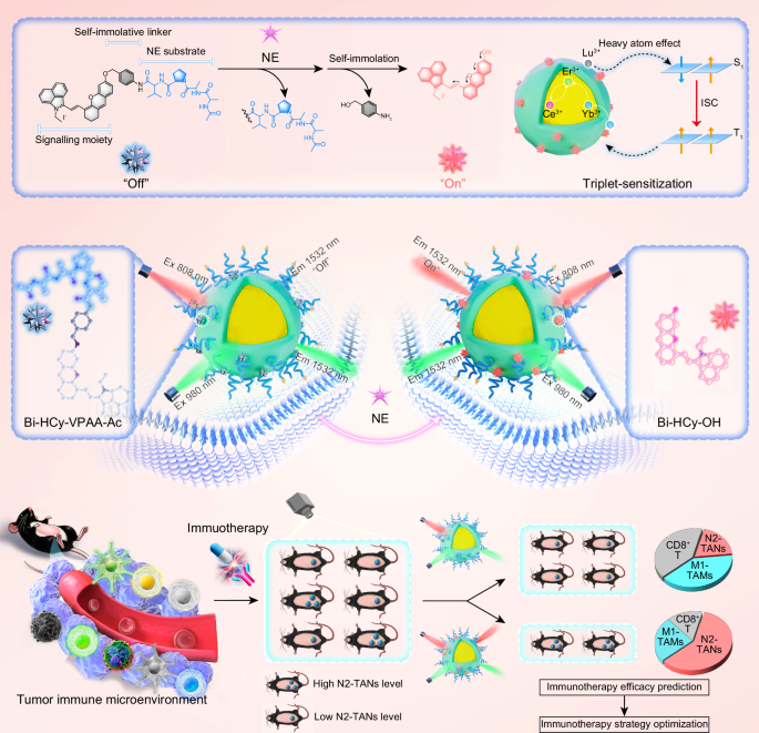
Synthesis and characterization of NE-responsive NIR-II fluorescence probes
The custom-synthesized NIR-II fluorescence dye, Bi-HCy-OH (benzo[cd]indole hemicyanine), was designed and synthesized through a series of reactions, utilizing commercial indole as the starting fluorophore (Supplementary Fig. 1). Then the Bi-HCy-VPAA-Ac was designed and synthesized between Bi-HCy-OH as a fluorophore and p-aminobenzyl alcohol-VPAA-Ac (pABA-VPAA-Ac) as a recognizer towards NE biomarker (Supplementary Fig. 1a). The chemical structures of Bi-HCy-OH, pABA-VPAA-Ac, and Bi-HCy-VPAA-Ac were verified by proton nuclear magnetic resonance (1H NMR) spectra, 13C NMR spectra, and mass spectrometry (MS) analysis (Supplementary Figs. 2–13 and Supplementary Data 1–25).
To explore the NE responsiveness of Bi-HCy-VPAA-Ac, we tested the absorption and fluorescence emission spectra of Bi-HCy-OH, Bi-HCy-VPAA-Ac, and Bi-HCy-VPAA-Ac plus NE. Upon NE addition, the absorption peak of Bi-HCy-VPAA-Ac red-shifted from ~665 nm to ~794 nm, mirroring the spectrum of Bi-HCy-OH (Fig. 2a). Compared to Bi-HCy-VPAA-Ac alone, the emission peak of Bi-HCy-VPAA-Ac plus NE significantly increased by 15.0-fold at ~962 nm, reaching levels comparable to Bi-HCy-OH (Fig. 2b). Furthermore, the conversion of Bi-HCy-VPAA-Ac into Bi-HCy-OH triggered by NE was proved by mass spectra (Fig. 2c–e, Supplementary Figs. 12–13, Supplementary Data 1-4, and Supplementary Data 21-25).

a Adsorption spectra, b fluorescence spectra, and (c, d) Mass spectra of pABA-VPAA-Ac, Bi-HCy-OH, Bi-HCy-VPAA-Ac, and Bi-HCy-VPAA-Ac cleaved by NE. e Illustration of proposed mechanism for cleavage and transformation of Bi-HCy-VPAA-Ac triggered by NE. f DFT calculation analysis of Bi-HCy-VPAA-Ac and Bi-HCy-OH. g TEM images, HRTEM images, FFT analysis, and SAED patterns of ErNPs and Bi-HCy-ErNPs. h HAADF-STEM images and STEM-EDX elemental mapping images of ErNPs, Bi-HCy-ErNPs without surface-functionalization of interpenetrating polymer network, and Bi-HCy-ErNPs. Experiments in (g) and (h) were repeated three times independently with similar results. i Hydrodynamic size distribution of ErNPs and Bi-HCy-ErNPs. j PXRD patterns of ErNPs and Bi-HCy-ErNPs. k Time-resolved fluorescence spectra of ErNPs and Bi-HCy-ErNPs.
To investigate the responsive mechanism of Bi-HCy-VPAA-Ac towards NE, we calculated the highest occupied molecular orbital (HOMO) and lowest unoccupied molecular orbital (LUMO) energy for Bi-HCy-OH and Bi-HCy-VPAA-Ac (Fig. 2f). The bandgap between the LUMO and HOMO of Bi-HCy-OH ( ~ 1.34 eV) is narrower compared to Bi-HCy-VPAA-Ac (~2.05 eV), suggesting that the electron donor (4-vinyl-2,3-dihydro-1H-xanthen-6-ol) in Bi-HCy-OH possesses a stronger “push-pull” electron system and electron-supplying capacity than that (1-(2-(2-acetamidopropanamido)propanoyl)-N-(3-methyl-1-oxo-1-((4-(((4-vinyl-2, 3-dihydro-1H-xanthen-6-yl)oxy)methyl)phenyl)amino)butan-2-yl)pyrrolidine-2-carboxamide) in Bi-HCy-VPAA-Ac. This could result in a stronger ICT effect to red-shift the absorption and emission peaks for Bi-HCy-OH compared to those for Bi-HCy-VPAA-Ac.
Synthesis and characterization of NIR-IIb fluorescnece ratiometric nanoprobes
To establish the FRET pathway, we initially adopted core-shell ErNPs, NaYbF4: Er, Ce@NaYF4: Yb, Lu, capable of energy transfer from dye to Yb3+ and subsequently to Er3+. Initially, NaYbF4: Er, Ce core nanocrystals were synthesized via a typical co-precipitation method, then the NaYF4: Yb, Lu shell was warped on the core surface by seed-mediated shell growth approach. Subsequently, the Bi-HCy-VPAA-Ac dye, with hydrophobic aromatic rings, was absorbed on the surface of ErNPs within a hydrophobic cavity created by oleic acid/1-octadecene ligands. Lastly, the cRGD-targeting and interpenetrating polymer network was further functionalized on the surface of dye-sensitized NaYbF4: Er, Ce@NaYF4: Yb, Lu through hydrophobic interactions and successive 1-(3-dimethylaminopropyl)−3-ethylcarbodiimide hydrochloride (EDC) chemistry between PMH/8Arm-PEG-NH2, 8Arm-PEG-NH2/PAA, and PAA/“DSPE-PEG-NH2/8Arm-PEG-NH2”, forming the final Bi-HCy-ErNPs nanoprobes. This surface functionalization could be expected to improve the nanoprobes’ stability and biocompatibility, enhance tumor accmulation, and protect the nanoprobes from degradation and aggregation, thus optimizing imaging performance. The synthesis of both ErNPs and Bi-HCy-ErNPs nanoparticles were verified by transmission electron microscope (TEM) images, high-resolution TEM (HRTEM) images, fast Fourier Transform (FFT) analysis, and selected area electron diffraction (SAED) patterns (Fig. 2g and Supplementary Fig. 14). Specifically, HRTEM images revealed distinct lattice stripes, with a lattice spacing of 2.18 Å for the core and 0.87 Å for the shell, proving the formation of core-shell structure of ErNPs. The high-angle annular dark field-scanning transmission electron microscopy (HAADF-STEM) and STEM-energy dispersive X-ray (STEM-EDX) elemental mapping images revealed the formation of ErNPs, with homogeneous distribution of Yb, Er, Ce, Lu, Y, Na, and F elements within the spatially defined regions, (Fig. 2h). Moreover, the introduction of both O and P within structure indicated the formation of Bi-HCy-ErNPs. The average hydrodynamic particle sizes of both ErNPs and Bi-HCy-ErNPs, determined by dynamic light scattering (DLS), were ~21.68 nm and ~29.69 nm, respectively (Fig. 2i). The powder X-ray diffraction (PXRD) patterns of both ErNPs and Bi-HCy-ErNPs presented their α-phase structure of NaYF4/NaYbF4 (Fig. 2j), which was conducive to efficient fluorescence emission at a low crystallographic symmetry. The fluorescence lifetime of Bi-HCy-OH and Bi-HCy-ErNPs was determined as ~1.43 and ~0.72 ns, respectively (Fig. 2k). This indicated the energy transfer from Bi-HCy-OH to ErNPs with an estimated energy transfer efficiency of ~49.65%. The X-ray photoelectron spectroscopy (XPS) verified the composition of Yb, Er, Ce, Lu, Y, Na, F, O, and P elements in the Bi-HCy-ErNPs nanoprobes (Supplementary Fig. 15).
To verify the feasibility of sensitization effect of Bi-HCy-OH towards ErNPs, we investigated their absorption and emission spectra. The absorption of ErNPs at 808 nm was appreciably weak, leading poor excitation efficiency at this wavelength (Fig. 3a). Consequently, we selected Bi-HCy-OH as a fluorophore for synthesizing NE-responsive Bi-HCy-VPAA-Ac, with the aim of sensitizing ErNPs. This selection was based on Bi-HCy-OH’s robust emission peak at ~962 nm (Fig. 3b), which aligned well with absorption peak of Yb3+ in ErNPs (Fig. 3c). This could help energy transfer from Bi-HCy-OH to Er3+ through Yb3+ underlying the FRET mechanism (Fig. 3d-f). With Lu3+-assisted dye-triplet-sensitization, ErNPs could emit intensified fluorescence at 1532 nm upon 808 nm excitation (Fig. 3f and Supplementary Fig. 16). Notably, the heavy atom Lu played a pivotal role in the formation of Bi-HCy-ErNPs, and its introduction into Bi-HCy-ErNPs was critical for intensifying fluorescence (Supplementary Fig. 16). After Bi-HCy-OH was absorbed onto the surface of ErNPs, the ErNPs exhibited a remarkable increase in fluorescence intensity when excited at 808 nm, but the fluorescence intensity remained constant when excited at 980 nm (Fig. 3g, h). This further proved the successful sensitization effect of Bi-HCy-OH towards ErNPs at 808 nm excitation, establishing a foundation for designing of NE-responsive NIR-IIb fluorescence ratiometric nanoprobes. Upon NE addition to Bi-HCy-VPAA-Ac, a newly red-shifted absorption peak emerged at ~794 nm, would be intensified, while the initial absorption peak at ~665 nm would be decreased (Fig. 3i). The emission peak at ~948 nm underwent a slight shift to ~962 nm, accompanied by a significant intensification (Fig. 3j). Consequently, upon NE triggering, the emission peak of Bi-HCy-ErNPs at 1532 nm increased significantly when excited at 808 nm, while keeping stable at 980 nm excitation (Fig. 3k-l). Moreover, this phenomenon exhibited dynamics dependent on both incubation time (Supplementary Fig. 17) and NE concentration (Fig. 3m, n). The NIR-II fluorescence ratiometric signals was linearly proportional to the NE concentration ranging from 0 to 1.6 μM (Fig. 3o, p), indicating that the Bi-HCy-ErNPs nanoprobes possessed high sensitivity for quantitative NE imaging, with a limit of detection (LOD, 3σ/slope) of ~0.37 ng/mL. Additionally, the nanoprobes demonstrated exceptional particle size stability, dye-loading stability, and optical stability in physiological environment including 10% FBS over time (Supplementary Figs. 18-22).

a Adsorption spectra of Bi-HCy-OH and ErNPs. b Fluorescence spectra of Bi-HCy-OH at 808 nm excitation and ErNPs at 980 nm excitation. c Absorption spectrum of ErNPs and fluorescence spectrum of Bi-HCy-OH at 808 nm excitation. d Proposed mechanism of Bi-HCy-VPAA-Ac and Bi-HCy-ErNPs in response to NE. e Schematic illustration of Bi-HCy-ErNPs in response to NE for NIR-IIb fluorescence ratiometric imaging. f Proposed dye-sensitization mechanism of Bi-HCy-OH towards ErNPs via energy transfer. g, h Fluorescence spectra of ErNPs and Bi-HCy-OH-ErNPs at (g) 808 and (g) 980 nm excitation. Dashed line indicated fluorescence emission peak. i Absorption spectra of Bi-HCy-VPAA-Ac before and after NE triggering. j Fluorescence spectra of Bi-HCy-VPAA-Ac before and after NE triggering at 808 nm excitation. k, l Fluorescence spectra of Bi-HCy-ErNPs before and after NE triggering at (k) 808 and (l) 980 nm excitation. Dashed line indicated fluorescence emission peak. m, n Fluorescence spectra of Bi-HCy-ErNPs triggered by NE at varying concentrations at (m) 808 and (n) 980 nm excitation. o Fluorescence intensity and (p) fluorescence ratiometric signal of Bi-HCy-ErNPs triggered by NE at varying concentrations at 808 and 980 nm excitation. Inset: NIR-IIb fluorescence imaging of Bi-HCy-ErNPs triggered by NE at varying concentrations under 808 and 980 nm excitation and their NIR-IIb fluorescence ratiometric imaging (n = 6).
Selective response of NIR-IIb fluorescence ratiometric nanoprobes towards NE
To assess the selective response of Bi-HCy-ErNPs nanoprobes towards NE, we employed the NE inhibitor (sivelestat), which molecular structure could compete with substrates to bind to the catalytic site of NE, to block their interactions36, thus effectively inhibiting NE activity. Meanwhile, the cathespin S, cathespin C, cathespin G, caspase-1, caspase-3, granzyme B, proteinase 3, cathepsin B, carboxylesterase, aminopeptidase N, leucine aminopeptidase, common ions (Na+, K+, Fe2+, and Ca2+), and some small molecules (glutathione and hydrogen peroxide) were taken as the controls. Then we conducted the NIR-IIb fluorescence spectroscopy and imaging studies (Fig. 4a). Upon exposure to the controls, minimal changes were observed in both F1532em, 808ex and F1532em, 980ex signals (Fig. 4b–g and Supplementary Figs. 23, 24). Consequently, no significant variation was observed in the ratio of F1532em, 808ex/F1532em, 980ex. In contrast, upon NE triggering, a significant increase was observed in the F1532em, 808ex signal, while the F1532em, 980ex signal remained stable, resulting in a significant increase in the ratio of F1532em, 808ex/F1532em, 980ex, and such ratio signal increase can be significantly abolished by adding the NE inhibitor. Furthermore, the increase in the F1532em, 808ex signal exhibited a concentration-dependent relationship with NE, as demonstrated by NIR-IIb fluorescence spectra (Fig. 4h and Supplementary Fig. 25). These findings suggested that Bi-HCy-ErNPs nanoprobes exhibited a highly selective response towards NE, enabling their effective recognition in complex biosystems.

a Schematic illustration of sensitivity and specificity of Bi-HCy-ErNPs in response to NE. b, c Fluorescence spectra of Bi-HCy-ErNPs treated with different formulations (NE inhibitor, cathespin S, cathespin C, cathespin G, caspase-1, caspase-3, granzyme B, and proteinase 3) at (b) 808 and (c) 980 nm excitation. d, e Fluorescence intensity of Bi-HCy-ErNPs treated with different formulations at (d) 808 and (e) 980 nm excitation (n = 50). Data are compared through one-way ANOVA with Tukey’s post-hoc test for multiple comparisons. d P-values of Bi-HCy-ErNPs + NE group to the other groups are <0.0001. e P-values of Bi-HCy-ErNPs + NE group to the other groups are >0.9150. f Fluorescence ratiometric signal of Bi-HCy-ErNPs treated with different formulations (n = 50). Data are compared through one-way ANOVA with Tukey’s post-hoc test for multiple comparisons. P-values of Bi-HCy-ErNPs + NE group to the other groups are <0.0001. g NIR-IIb fluorescence imaging of Bi-HCy-ErNPs treated with different formulations at 808 and 980 nm excitation and their NIR-IIb fluorescence ratiometric imaging. h Fluorescence spectra of Bi-HCy-ErNPs treated with different formulations at varying concentrations at 808 nm excitation.
In vitro TANs imaging using NIR-IIb fluorescence ratiometric nanoprobes
Motivated by in vitro imaging ability of Bi-HCy-ErNPs towards NE, we investigated the efficacy of Bi-HCy-ErNPs nanoprobes in detecting endogenous NE in different types of cells, including N1-neutrophils, N2-neutrophils, CD8+ T cells (CD8+ Ts), M1-bone marrow derived macrophages (M1-BMDM), NIH/3T3, and Hepa1-6 cell. Prior to in vitro imaging, we initially assessed the biocompatibility of Bi-HCy-ErNPs nanoprobes by standard 3-(4, 5-dimethyl-2-thiazolyl)-2, 5-diphenyl-2-tetrazolium bromide method. After incubating various cell types with nanoprobes for 24 h, we observed over 80% cell viability, even at the concentration of up to 1000 μg/mL, indicating the excellent biocompatibility of Bi-HCy-ErNPs (Supplementary Figs. 26, 27). Next, we utilized the nanoprobe to detect intracellular NE levels. Various cell lines were incubated in a 96-well plate and further incubated with Bi-HCy-ErNPs nanoprobes for 8 h. Subsequently, the NIR-IIb fluorescence images and signals at 1532 nm were recorded upon 808 and 980 nm excitation. The results showed that the N2-neutrophils emitted strong F1532em, 808ex signals as well as robust F1532em, 980ex signals, concurrently accompanied by a high ratiometric signal (Fig. 5a–c). In contrast, other cell types, when incubated with Bi-HCy-ErNPs over identical time intervals, manifested weak F1532em, 808ex signals, yet still presented strong F1532em, 980ex signals, along with a relatively low ratiometric signal. Furthermore, pre-exposure to the NE inhibitor sivelestat (100 μM) significantly decreased the NIR-IIb fluorescence of N2-neutrophils incubated with Bi-HCy-ErNPs at 808 nm excitation, whereas the fluorescence remained stable at 980 nm excitation (Fig. 5a–c). Consequently, the ratiometric signal was significantly decreased. These results indicated high NE-responsive selectivity of Bi-HCy-ErNPs nanoprobes towards N2-neutrophils17,27,37. Additionally, the bio-TEM imaging revealed that N2-neutrophils incubated with Bi-HCy-ErNPs effectively internalized the probes and then distributed them within intracellular spaces (Fig. 5d).

a NIR-IIb fluorescence ratiometric imaging of different types of cells cultured within plates for 12 h and incubated with Bi-HCy-ErNPs for 8 h. b Fluorescence spectra of N1-neutrophils and N2-neutrophils incubated with Bi-HCy-ErNPs for 8 h. c NIR-IIb fluorescence ratiometric quantification/ratiometric analysis of different types of cells cultured within plates for 12 h and incubated with Bi-HCy-ErNPs for 8 h (n = 50). d Bio-TEM images of N2-neutrophils incubated with Bi-HCy-ErNPs for 8 h in culture mediums. e NIR-IIb fluorescence ratiometric imaging of different types of cells incubated with Bi-HCy-ErNPs for 8 h. Experiments in (d) and (e) were repeated three times independently with similar results. f NIR-IIb fluorescence quantification/ratiometric analysis of different types of cells incubated with Bi-HCy-ErNPs for 8 h (n = 50). g NIR-IIb fluorescence ratiometric imaging and (h) ratiometric analysis of different types of cells cultured within plates for 12 h and incubated with Bi-HCy-ErNPs after 0.5, 1, 2, and 4 h (n = 50). i Correlation between fluorescence intensity of antibodies towards leukocytes’ biomarkers and NIR-IIb fluorescence ratiometric signal via a simple linear regression model in Bi-HCy-ErNPs-incubated leukocytes. Centre line in black shows the best-fit linear regression, and error band in gray shows 95% confidence intervals of linear regression line (n = 6). R and ρ values were derived using a simple linear regression model. Data are presented as mean ± standard deviation (c−h). Data are compared through one-way ANOVA with Tukey’s post-hoc test for multiple comparisons (c−h). P-values (N2-neutrophils group vs. the other groups) are <0.0001, >0.6190, and <0.0001 in F808ex, F980ex, and ratiometric signal detection (c). P-values (N2-neutrophils group vs. the other groups) are <0.0001, >0.0570, and <0.0001 in F808ex, F980ex, and ratiometric signal detection (f). P-values (0.5 h vs. 1 h, 1 h vs. 2 h, and 2 h vs. 4 h) are 0.5874, <0.0001, and <0.0001, respectively (h).
The effectiveness of Bi-HCy-ErNPs nanoprobes was further investigated by using NIR-IIb fluorescence microscopy. The F1532em, 980ex signals exhibited similarity across all cells, whereas the F1532em, 808ex signals and ratiometric signals were significantly elevated in the N2-neutrophils compared to the N1-neutrophils, NE inhibitor-treated N2-neutrophils, and other cells (Fig. 5e, f). The result was likely attributable to the interaction between Bi-HCy-ErNPs and intracellular NE, which sensitized ErNPs through the time-dependent formation of Bi-HCy-OH molecules (Fig. 5g, h and Supplementary Fig. 28). After assessment using a simple linear regression model, a remarkably strong positive correlation was found between NIR-IIb fluorescence ratiometric signal of Bi-HCy-ErNPs and specific dye-labeled antibodies towards cellular biomarkers with a correlation coefficient (R) 0.84-0.98 and a Pearson’s r value (ρ) of 0.91-0.96 (Fig. 5i). The data further proved the fluorescence activation of Bi-HCy-ErNPs required NE biomarkers from N2-neutrophils. Collectively, these results demonstrated that Bi-HCy-ErNPs nanoprobes had the potential to serve as reporters for distinguishing cells with varying NE levels, which might enable quantitative imaging of TANs with high specificity.
In vivo TANs visualization using NIR-IIb fluorescence ratiometric nanoprobes
Encouraged by the nanoprobe’s in vitro exceptional performance for precise NIR-IIb fluorescence imaging towards NE, we conducted further investigations in Hepa1-6 tumor-bearing C57BL/6 mice and H22 tumor-bearing BALB/c mice38,39,40. Prior to in vivo imaging, we initially assessed the blood compatibility of Bi-HCy-ErNPs nanoprobes. The appreciably low hemolysis rate indicated that Bi-HCy-ErNPs nanoprobes were applicable to intravenous administration (Supplementary Fig. 29). When the tumor grew to a certain volume, the tumor-bearing mice received immunotherapy on day 2, 4, and 6 followed by intravenous injection of nanoprobe through the tail vein on day 7 (Fig. 6a). Whole-body NIR-IIb fluorescence imaging at 1532 nm was recorded at various time points upon excitation at 808 and 980 nm. Upon excitation at 980 nm, a strong and persistent NIR-IIb fluorescence signal was observed in the tumors of both non-treatment and the treatment groups (Fig. 6b), which was attributed to the integrated passive and active tumor-targeting of nanoprobes41 with their prolonged circulation lifetime (Fig. 6b, and Supplementary Figs. 30–33). Conversely, the tumors of the treatment group emitted significantly weaker NIR-IIb fluorescence signals compared to non-treatment group upon excitation at 808 nm. Moreover, upon intratumor injection of NE inhibitor into non-treatment groups, the NIR-IIb fluorescence of Bi-HCy-ErNPs nanoprobes at the tumor sites was significantly declined when excited at 808 nm, but remained relatively stable at 980 nm excitation, indicating the selectivity of Bi-HCy-ErNPs nanoprobes towards NE in vivo. Quantitative analysis revealed that the ratiometric signals of tumor regions in vivo in the treatment group were ~8.0-fold higher compared to those in non-treatment group (Fig. 6c). NIR-IIb fluorescence imaging of tumor lysate also showed stronger ratiometric signals in the treatment group as opposed to non-treatment group (Fig. 6d, e), aligning with the in vivo NIR-IIb fluorescence imaging results. These results were further confirmed by the gold standard hematoxylin and eosin (H&E) staining (Supplementary Figs. 34–35). In addition, body weight assessment, H&E staining, blood biochemical assay, and hematological indexes exhibited that intravenous administration of Bi-HCy-ErNPs did not lead to apparent histological abnormalities/lesions and had no pronounced inflammation/infection, nephrotoxicity, hepatotoxicity, or hematological toxicity (Supplementary Figs. 36–38). We also investigated biodistribution and clearance profiles of Bi-HCy-ErNPs by inductively coupled plasma optical emission spectrometry (ICP-OES). It was observed that the nanoprobes from urine and feces reached their maximum at the 4th day and gradually decreased to normal levels over 14 days (Supplementary Fig. 39). Moreover, ~85% of injected nanoprobes were excreted from the body within 14 days (Supplementary Fig. 40). This indicated that the nanoprobes could be cleared from the body through both hepatobiliary and renal excretion pathways over time. Regarding long-term potential immune responses, the key cytokines like pro-inflammatory cytokines (TNF-α, IL-12, IL-6, and IFN-γ) and anti-inflammatory cytokine (IL-10) showed no significant changes between nanoprobe-injected and PBS-injected groups (Supplementary Figs. 41–43), indicating Bi-HCy-ErNPs caused no abnormal cytokine secretion, reducing adverse immune responses. Additionally, common immune cells revealed no significant population changes after nanoprobe use (Supplementary Fig. 44). This implied that Bi-HCy-ErNPs maintained immune cell homeostasis during long-term use. Overall, these results demonstrated the excellent in vivo biosafety profile of Bi-HCy-ErNPs nanoprobes, supporting their potential for clinical translation. To investigate the correlation between activatable signals of nanoprobes and intratumoral neutrophils, we performed immunofluorescence staining at the end of imaging process. The NIR-IIb fluorescence signal of Bi-HCy-ErNPs in tumor sections from non-treatment group excited at 808 nm was 10.2-fold higher stronger than in those from the treatment group, and showed a high co-localization of ~81.4% with NE-antibody (YA5765, MedChemExpress, USA)-labeled TANs (Fig. 6f, g). Clinical data have demonstrated that elevated levels of M1-tumor-associated macrophages (M1-TAMs) and CD8+ Ts were closely correlated with a favorable therapeutic prognosis42,43. In contrast, an increase in N2-TANs was strongly indicative of lower survival rates among cancer patients43,44. Thus, the trend of immune status changes reflected by the level variations of M1-TAMs/CD8+ Ts was opposite to that of N2-TANs. As depicted in Fig. 6f–l, the change in immunofluorescence signals of N2-TANs following immunotherapy treatment was diametrically contrary to that of M1-TAMs and CD8+ Ts, which further validated the high accuracy of our N2-TANs imaging.

a Illustration of imaging and immunotherapy schedule. b NIR-IIb fluorescence ratiometric imaging and (c) NIR-IIb fluorescence quantification/ratiometric analysis of different groups. In the immunotherapy group, the mice received intraperitoneal injection of anti-PD-L1 antibody on alternate days. On day 7, the mice received intravenous injection with Bi-HCy-ErNPs (n = 6). d NIR-IIb fluorescence ratiometric imaging of tumor lysate from tumor-bearing mice without/with immunotherapy after intravenous injection of Bi-HCy-ErNPs. e NIR-IIb fluorescence quantification/ratiometric analysis of tumor lysate from tumor-bearing mice without/with immunotherapy after intravenous injection of Bi-HCy-ErNPs (n = 50). f Co-localization of NIR-IIb fluorescence signals of Bi-HCy-ErNPs excited at 808 nm with N2-TANs/M1-TAMs/CD8+ Ts in tumor sections. Venn diagram indicated the average cell numbers, and the table indicated the percentages of single- and double-positive cells of Bi-HCy-ErNPs excited at 808 nm and N2-TANs/M1-TAMs/CD8+ Ts. g−i Ex vivo immunofluorescence imaging and (j-l) NIR-IIb fluorescence quantification/ratiometric analysis of tumors from tumor-bearing mice without/with immunotherapy after intravenous injection of Bi-HCy-ErNPs (n = 16). False-blue fluorescence indicated the signal of nucleus. False-green fluorescence indicated the signal of (g, j) N2-TANs, h, k M1-TAMs, and (i, l) CD8+ Ts, labeled by different antibodies. False-yellow and false-cycan fluorescence indicated the signal of Bi-HCy-ErNPs at 808 and 980 nm excitation. Dashed line indicated the basic outline of a mouse model. Data are presented as mean ± standard deviation (c−l). Data are compared with two-tailed unpaired Student’s t-test (e−l). P-values (non-treatment vs. treatment) are <0.0001, 0.9651, and <0.0001 in F808ex, F980ex, and ratiometric signal detection (e). P-values (non-treatment vs. treatment) are <0.0001, 0.1934, and <0.0001 in F808ex, F980ex, and ratiometric signal detection (j). P-values (non-treatment vs. treatment) are <0.0001, 0.1643, and <0.0001 in F808ex, F980ex, and ratiometric signal detection (k). P-values (non-treatment vs. treatment) are <0.0001, 0.5480, and <0.0001 in F808ex, F980ex, and ratiometric signal detection (l).
In vivo TANs visualization using NIR-IIb fluorescence ratiometric nanoprobes for immunotherapy efficacy monitoring
To explore the potential of Bi-HCy-ErNPs nanoprobe in immunotherapy efficacy monitoring, we conducted the prolonged NIR-IIb fluorescence ratiometric imaging (Fig. 7a). As illustrated in Fig. 7b, c, on day 8, the NIR-IIb fluorescence ratiometric signals in the treatment group surged abruptly, indicating that the immunotherapy started to produce effects, leading to a change in the immune status of TANs and a significant decrease in NE expression. The results were corroborated by the H&E staining of tumor slices (Supplementary Fig. 45). We further carried out long-term NIR-IIb fluorescence ratiometric imaging and immunotherapy efficacy monitoring (Fig. 7d). As depicted in Fig. 7e, f, the treatment group led to markedly intensified NIR-IIb fluorescence ratiometric signals, while the NIR-IIb fluorescence ratiometric signals remained unaltered in non-treatment group. In addition, we noticed a negative correlation between the NIR-IIb fluorescence ratiometric signal and tumor volumes/weights in Hepa1-6 tumor-bearing mice (Fig. 7g, h), which was consistent with the H&E staining of tumor slices (Supplementary Fig. 46). Moreover, the population of TANs, TAMs, CD8+ Ts, mature dendritic cells (mDCs), tumor-infiltrating effector memory T cells (Tems), and tumor-infiltrating regulatory T cells (Tregs) was further quantified by flow cytometry at the end of imaging (Fig. 7i, j, and Supplementary Figs. 47–51). The proportion of M1-TAMs, CD8+ Ts, mDCs, and Tems in the treatment group was significantly increased compared to that in the non-treatment mice. Additionally, there was a significant decrease in the proportion of M2-TAMs and Tregs in the treatment group, in contrast to the non-treatment group. Notably, compared to the non-treatment group, the number of TANs in the treatment group was markedly reduced to ~5.54%, nearly 4.24-fold lower than the non-treatment group (Fig. 7i). The results indicated a robust correlation between the NIR-IIb fluorescence ratiometric signals of Bi-HCy-ErNPs and TANs’ immune status changes, suggesting its promising potential as a tool for assessing immunotherapeutic responses.

a Illustration of long-term imaging and immunotherapy schedule (n = 6 mice for non-treatment and treatment group, respectively). b NIR-IIb fluorescence ratiometric imaging and (c) NIR-IIb fluorescence quantification/ratiometric analysis of tumor-bearing mice without/with immunotherapy after intravenous injection of Bi-HCy-ErNPs. d Illustration of long-term imaging and immunotherapy schedule. e NIR-IIb fluorescence ratiometric long-term imaging and (f) NIR-IIb fluorescence quantification/ratiometric analysis of tumor-bearing mice without/with immunotherapy after intravenous injection of Bi-HCy-ErNPs (n = 6 mice). g Tumor growth curves, excised tumor weight, and body weight change of tumor-bearing mice without/with immunotherapy (n = 6 mice). Absolute tumor volume measured for each group is included in Source Data file. h Correlation between NIR-IIb fluorescence ratiometric signal and tumor volumes of tumor-bearing mice with immunotherapy via a simple linear regression model. Centre line in black shows the best-fit linear regression, and error band in gray shows 95% confidence intervals of linear regression line. i Flow cytometric assay of TANs at the end of imaging. j Flow cytometric assay of mDCs (CD11c+CD80+CD86+) in tumor-draining lymph nodes (TDLNs), Tems (CD3+CD8+CD62LlowCD44hi) in spleen, tumor-infiltrating CD8+ Ts in CD3+ Ts (CD3+CD8+), M1-TAMs (CD11b+F4/80+CD80hi), M2-TAMs (CD11b+F4/80+CD206hi), and tumor-infiltrating Tregs (CD4+Foxp3+) (n = 6 mice). Dashed line indicated the basic outline of a mouse model. Data are presented as mean ± standard deviation (c−j). Data are compared through one-way ANOVA with Tukey’s post-hoc test for multiple comparisons (c and f) and two-tailed unpaired Student’s t-test (g and j). P-value (0/1/4 d vs. 5/8/9/12/13 d) is <0.0001 in F808ex detection (c). P-value (0/1/4/5 d vs. 8/9/12/13 d) is <0.0001 in ratiometric signal detection (c). P-values (0 d vs. 7/14/21 d) are <0.0001 in F808ex and ratiometric signal detection (f). P-values (non-treatment vs. treatment) in tumor volume and tumor weight measurement are <0.0001 (g). P-values (non-treatment vs. treatment) in are <0.0001 in mDCs, Tems, CD8+ Ts, M1-TAMs, M2-TAMs, and Tregs detection (j).
In vivo TANs visualization using NIR-IIb fluorescnece ratiometric nanoprobes for immunotherapy efficacy monitoring, individual stratification, and immunotherapy strategy optimization
To assess the potential of Bi-HCy-ErNPs nanoprobe in individual stratification, we established Hepa1-6 tumor-bearing C57BL/6 and H22 tumor-bearing BALB/c mouse models to distinguish between responder and non-responder to immunotherapy via NIR-IIb fluorescence ratiometric imaging (Fig. 8a). Initially, nine C57BL/6 mice were inoculated with Hepa1-6 cells to establish Hepa1-6 tumor-bearing C57BL/6 mice. After tumor reached a certain range of volume, all nine mice underwent immunotherapy on days 0, 2, and 4. On day 20, all nine mice received an intravenous injection of Bi-HCy-ErNPs. Subsequently, we assessed NE responsiveness at 24 h post-injection using NIR-IIb fluorescence ratiometric imaging. As depicted in Fig. 8b-d, six mice (mice 4-9) were categorized into “responder group” based on robust NIR-IIb fluorescence ratiometric signals due to high NE levels within tumor microenvironment. Conversely, the remaining three mice were assigned to “non-responder group” owing to feeble NIR-IIb fluorescence ratiometric signals, which could be ascribed to relatively low NE levels within tumor microenvironment. In addition, we observed a negative correlation between the NIR-IIb fluorescence ratiometric signal and tumor volumes/weights/NE expression levels in Hepa1-6 tumor-bearing mice as well (Fig. 8e, f and Supplementary Fig. 52). Furthermore, the results obtained by H&E (hematoxylin and eosin)/TUNEL(terminal deoxynucleotidyl transferase dUTP nick end labeling)/Ki-67 staining as well as TANs/CD8+ Ts staining of tumor slices from Hepa1-6 tumor-bearing mice, proved the distinctions in cellular damage between NE-responsive and NE-irresponsive groups upon immunotherapy (Fig. 8g). These findings revealed significant differences in TAN immune status between NE-responsive and NE-nonresponsive groups, which could be visualized in real-time using Bi-HCy-ErNPs nanoprobes. In addition, NE is primarily secreted by neutrophils, and its expression level is closely associated with tumor invasion and metastasis. No systematic classification was established between NE expression levels and specific tumor types9,45,46,47,48,49. Thus, the similar results were also observed in the H22 and 4T1 tumor-bearing BALB/c mice (Fig. 8b-e, f, g, and Supplementary Figs. 53-55).

a Illustration of immunotherapy and imaging schedule (n = 9 mice for each group, respectively). b NIR-IIb fluorescence ratiometric imaging of both immunotherapy-irresponsive and immunotherapy-responsive individuals after intravenous injection of Bi-HCy-ErNPs. c NIR-IIb fluorescence quantification/ratiometric analysis of both immunotherapy-irresponsive and immunotherapy-responsive individuals after intravenous injection of Bi-HCy-ErNPs (n = 3 and 6 mice for non-responder and responder group, respectively). d Illustration of in vivo NIR-IIb fluorescence ratiometric imaging for individual stratification. e Tumor growth curves and excised tumor weight of both immunotherapy-irresponsive and immunotherapy-responsive individuals with immunotherapy (n = 9, 3, and 6 mice for PBS, non-responder, and responder group, respectively). Absolute tumor volume measured for each group is included in Source Data file. f Correlation between NIR-IIb fluorescence ratiometric signal and tumor volumes of tumor-bearing mice with immunotherapy via a simple linear regression model. Centre line in black shows the best-fit linear regression, and error band in gray shows 95% confidence intervals of linear regression line. R and ρ values were derived using a simple linear regression model (n = 3 and 6 mice for non-responder and responder group, respecrively). g H&E/TUNEL/Ki-67 staining and TANs/CD8+ Ts staining in tumor slices from both immunotherapy-irresponsive and immunotherapy-responsive individuals with immunotherapy. Experiments were repeated three times independently with similar results. Dashed line indicated the basic outline of a mouse model. Data are presented as mean ± standard deviation (c and e). Data are compared with two-tailed unpaired Student’s t-test (c) and one-way ANOVA with Tukey’s post-hoc test for multiple comparisons (e). P-values (non-responder vs. responder) in F808ex, F980ex, and ratiometric signal detection from Hepa1-6 tumor-bearing mice are <0.0001, 0.5472, and <0.0001, respectively (c). P-values (non-responder vs. responder) in F808ex, F980ex, and ratiometric signal detection from H22 tumor-bearing mice are <0.0001, 0.4665, and 0.0002, respectively (c). P-values (non-responder vs. responder) in tumor volume and tumor weight measurement are <0.0001 (e).
Immune checkpoint inhibitors played major roles in immune escape by inhibiting immune responses and promoting immune tolerance toward tumor cells. Among these, PD-L1 and CTLA-4 have been identified as promising targets. The combined blockade of Immune checkpoint inhibitors might provide potentially synergistic effects due to the differences in the timing and location of their reactions. Therefore, in cases where monotherapy with anti-PD-L1 antibody (HY-P99145, MedChemExpress, USA) showed suboptimal efficacy, combination therapy with anti-CTLA-4 antibody (HY-P99132, MedChemExpress, USA) might achieve enhanced immunotherapeutic effects. Additionally, lenvatinib, a VEGFR inhibitor, has been shown to provide potential synergistic effects when used in conjunction with immune checkpoint inhibitors by inhibiting angiogenesis and reversing immunosuppressive tumor microenvironment. To further assess the potential of Bi-HCy-ErNPs nanoprobe in personalized immunotherapy, we delved deeper into immunotherapy efficacy monitoring, individual stratification, and immunotherapy strategy optimization (Fig. 9a). First, fifteen C57BL/6 mice were inoculated with Hepa1-6 cancer cells. Once tumor grew to a certain range of volume, all fifteen mice underwent immunotherapy via intraperitoneal injection of anti-PD-L1 antibody on days 0, 2, and 4. On day 13, an intravenous injection of Bi-HCy-ErNPs was administered to all the mice. Subsequently, individual stratification was carried out at 24 h post-injeciton, employing NIR-IIb fluorescence ratiometric imaging. As presented in Fig. 9b–e, nine mice (mice 7−15) were classified as responder group, whereas the remaining six mice (mice 1−6) were designated to non-responder group. Moreover, a negative correlation between the NIR-IIb fluorescence ratiometric signal and tumor volumes/weights was detected across all groups (Fig. 9e, f). Then, the non-responder group (mice 1-6) were further randomly divided into two groups including adjustor group (mice 1-3) and non-adjustor group (mice 4-6). Among the adjustor group, 3 mice further received immunotherapy by receiving intraperitoneal injection of anti-PD-L1 antibody plus anti-CTLA-4 antibody50 at the day 14, 16, and 18. At the day 20 and 27, the mice further received intravenous injection with Bi-HCy-ErNPs for imaging on days 21 and 28 to monitor immunotherapy efficacy. As illustrated in Figs. 9b-d, g, h and Supplementary Fig. 56, the in vivo NIR-IIb fluorescence ratiometric signals showed a gradual increase from day 14, via day 21, to day 28, suggesting an effective therapeutic response after adjusting the immunotherapy regimen. Furthermore, the comprehensive results, including ex vivo NIR-IIb fluorescence ratiometric imaging, TANs/CD8+ Ts staining, tumor evolution monitoring, flow cytometric assay of TANs/TAMs/CD8+ Ts, as well as H&E/TUNEL/Ki-67 staining of tumor slices from Hepa1-6 tumor-bearing mice (Fig. 9i–o), further validated the enhanced therapeutic efficiency after optimizing immunotherapeutic interventions. It also clearly demonstrated that the NIR-IIb fluorescence ratiometric imaging based on Bi-HCy-ErNPs probe could reliably identify the responders to immunotherapy. Then, the effectiveness of immunotherapy could be assessed in advance, which would in turn provide the possibility for timely adjustment of the immunotherapy strategy. This supported its use in guiding drug selection for precision medicine. Overall, our designed probe offered a promising visualization tool to monitor in vivo immune status change of TANs with high molecular imaging sensitivity. More importantly, our work held the potential to further expand into individual stratification so as to customize personalized treatment plan, with the aim of improving therapeutic efficacy in cancer immunotherapy.

a Illustration of optimized immunotherapy and imaging schedule (n = 15 mice for each group, respectively). b NIR-IIb fluorescence ratiometric imaging of non-responder and responder group on day 14. c, d NIR-IIb fluorescence ratiometric imaging of adjustor group on day (c) 21 and (d) 28. e NIR-IIb fluorescence ratiometric analysis of different groups on day 14 (n = 15, 6, and 9 mice for PBS, non-responder, and responder group, respectively). f Correlation between NIR-IIb fluorescence ratiometric signal and relative tumor volumes via a simple linear regression model. Centre line in black shows the best-fit linear regression, and error band in gray shows 95% confidence intervals of linear regression line. R and ρ values were derived using a simple linear regression model. g NIR-IIb fluorescence quantification and (h) NIR-IIb ratiometric analysis of adjustor group (n = 3 mice). i Ex vivo NIR-IIb fluorescence ratiometric analysis of tumors from different groups. j Tumor growth curves, k excised tumor weight, l body weight changes, and (m) survival rates of different groups (n = 15, 3, 3, and 9 mice for PBS, non-adjustor, adjustor, and responder group, respectively). Absolute tumor volume measured for each group is included in Source Data file. n Flow cytometric assay of TANs, TAMs, and CD8+ Ts from different groups. o H&E/TUNEL/Ki-67 staining and TANs/CD8+ Ts staining of tumors from different groups. Experiments were repeated three times independently with similar results (i and o). Data are presented as mean ± standard deviation (e−l). Data are compared through one-way ANOVA using Tukey’s post-hoc test (e−k), Dunnett’s comparison test (j), and log-rank (Mantel-Cox) test (m). P-values (non-responder vs. responder) in F808ex, F980ex, and ratiometric signal detection are <0.0001, 0.9699, and <0.0001 (e). P-values (day 21 vs. day 28) in F808ex, F980ex, and ratiometric signal detection are 0.0141, 0.8477, and 0.0016 (g, h). P-values (non-adjustor vs. responder) are 0.0103 (j), <0.0001 (k), and 0.0002 (m).