
Mice and cell culture
Female Balb/c mice (Jackson Laboratories) were housed and maintained at City of Hope. Mice were acclimatized for a minimum of 3 days prior to experimental procedures. Animal study protocols were approved by the Institutional Animal Care and Use Committee (IACUC) at City of Hope. Humane endpoints for euthanasia prior to pre-determined experimental endpoints were per institutional guidelines and included maximum tumor diameter >15 mm or tumor necrosis >80% of tumor surface area. Since this was a proof-of-concept study, sample size was determined by the number of mice that could be feasibly treated with RT or imaged in a single day. Mice were randomly allocated to unblinded treatment groups, stratified for size-matched tumors. For localized irradiation studies, sulfatrim feed or water was provided starting a few days prior to radiation and for at least 2 weeks after; hydrogel was provided post-irradiation. Animals were monitored at least 2–3 times per week (more frequently near endpoints) for radiation sickness, body condition, and tumor size.
4T1 tumor cells were obtained from ATCC (CRL-2539) and cultured per manufacturer’s instructions in Roswell Park Memorial Institute (RPMI, Corning Cat # 10-041-CV) supplemented with 10% fetal bovine serum (Corning Cat # 35-011-CV). Cells were passaged no more than 6 times prior to orthotopic injections and were regularly tested for Mycoplasma contamination.
Orthotopic tumor implantation and treatment
For orthotopic injections, tumor cells were suspended in 50 μl of Matrigel matrix (Corning Cat # 356237) and Hanks’ Balanced Salt Solution (HBSS, Corning Cat # 21-022-CV). Anti-CTLA4 (InVivoMab 9H10, Bio X Cell) was diluted in phosphate-buffered saline (PBS, Corning Cat # 21-031-CV). To generate size-matched tumors for untreated mice at the imaging timepoint, orthotopic injections were performed 6 days prior to radiotracer injection for the untreated cohort.
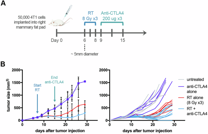
50 ×103 4T1 tumor cells were injected into the right fourth mammary fat pad of anesthetized 6–8 week-old female BALB/c mice; treatment began after 6 days when tumors were ~5 mm in diameter. Tumor growth was monitored by caliper measurement ((length of longest dimension) * (perpendicular width)2)/2.
For RT, tumors were treated conformally with 8 Gy fractions for 3 consecutive days to a total of 24 Gy. Radiation was delivered with a 1 cm collimator to anesthetized mice on the X-RAD SmART system using CT-based treatment planning. One day after completing RT, mice started treatment with anti-CTLA4 antibody; 200 µg was injected intraperitoneally every 3 days for a total of 3 doses1.
Anesthesia and euthanasia
Inhaled isoflurane up to 5% was used for anesthesia during tumor implantation, imaging, and radiation delivery. Depth was monitored by respiratory rate/color and loss of toe-pinch reflex. Euthanasia methods included CO₂ from compressed gas cylinders and/or isoflurane overdose, followed by a secondary physical method (bilateral thoracotomy or cervical dislocation) to confirm death.
Immunohistochemistry (IHC)
Tumors were harvested at multiple timepoints for analysis by hematoxylin and eosin (H&E) staining and IHC: 1 day after anti-CTLA4 x 3, 1 day after 8 Gy x 1, 1 day after 8 Gy x 3, 1 day after 8 Gy x3 + anti-CTLA4 x 1, 1 day after 8 Gy x 3 + anti-CTLA4 x 3, 5 days after 8 Gy x 3 + anti-CTLA4 x 3, 14 days after 8 Gy x 3 + anti-CTLA4 x 3. Except where indicated, control mice were treated according to the same schedule as mice treated with RT + anti-CTLA4, such that RT alone controls were harvested 8 days after completing RT, and anti-CTLA4 alone controls were harvested 1 day after the last dose of anti-CTLA4. For Visiopharm analysis, non-tumor adipose tissue was excluded by an independent reviewer, when present, to accurately characterize intratumoral lymphocytes.
Tumors were fixed in formalin and paraffin-embedded for IHC. CD8+ cell density on central tumor slices (cell count/mm2) was quantified with Visiopharm software using the same protocol across all samples. Two-dimensional cell density was calculated as (CD8+ cells for an entire tumor slice)/(total tumor area). Antibodies: anti-CTLA4 (InVivoMab 9H10, Bio X Cell), CD8α (D4WD2Z, Cell Signaling), CD4 (D7D2Z, Cell Signaling) and CD3 (2GV6, Ventana).
CD8 immunoPET imaging
Purified CD8α-specific cys-diabody was lightly reduced, conjugated with maleimide-desferrioxamine and radiolabeled with 89Zr as previously described10. Approximately 10 µg (3.7 MBq) of radiolabeled CD8 cys-diabody (89Zr-Df-CD8cDb) was administered by tail vein injection to each mouse 15 days after orthotopic injection of tumors treated with RT. Static PET/CT images were acquired 24–48 h after radiotracer injection. Images were acquired on a GNEXT small animal PET/CT scanner (SofieBiosciences). Imaging analysis was performed with Velocity software (V4.1, Varian Medical Systems, Inc. Palo Alto, CA).
Average %injected dose/cc (%ID/cc) was determined for the entire tumor contoured on axial CT images with treatment group blinded. For biodistribution studies, mice were euthanized after imaging, and tissues were dissected, weighed, and counted in a gamma counter.
Statistics
Comparisons for cell density by IHC were made by Kruskal-Wallis ANOVA with p-values adjusted for multiple comparisons, or unpaired two-tailed t-test when comparing two groups. Unpaired two-tailed t-test was used for comparisons of %ID/cc or %ID/g for imaging and biodistribution studies. Pearson correlation coefficient was computed for assessing linear correlation between %ID/cc for imaging and tumor volume or CD8+ cell density by IHC. GraphPad Prism v7.0 was used for statistical analysis. N-way analysis of variance (ANOVA) at each timepoint was used to evaluate statistical significance of tumor regression including interaction with CTLA4 (MATLAB, The MathWorks Inc., Natick, Massachusetts). Three treatment groups were considered: (1) CTLA4 alone, (2) RT alone, (3) CTLA4 + RT.